
Introduction
Globally, the Kenyan flower industry is the third major flower exporter by value and volume just behind the Netherlands and Colombia on a global level1 while in Africa, it is the largest producer and the leading supplier of Fair trade flowers to Europe accounting for 50-60% of total volumes.2 The flower production in the country is mainly large scale and export oriented.3 Cut flower is one of the two commodities where large-scale production dominates in the country and roses make up over 70% of all buds, trees, roots, flowers and foliage that is exported from Kenya.4 Roses alone account for about 35% of the total value of horticultural exports and therefore form the most important export product.5
In commercial production of roses, yield is restricted by an assortment of variables such as light, humidity, mineral nutrition, temperature, salinity and biotic factors.6 The common biotic factors in cut-flower roses include pests and diseases. Crown gall caused by A. tumefaciens causes a significant damage to roses in Kenya. The bacterium is found in the soil and it is responsible for the tumorous growth found in infected plants.7 The pathogen is widespread, naturally occurring soil bacterium that causes crown gall in numerous plant species in nurseries, commercial production and uncultivated areas and has the capacity to introduce new genetic material into the plant cell. Significant annual losses of between 5-6% are faced by the flower growers worldwide due to the disease in the form of lack of vigor, reduction in foliage, low quality rose flower, low productivity and increased susceptibility of infected plants to pathogens and environmental stress.8 In other instances, losses of between 10-30% in nursery stalk have been reported in fruit trees. Both yield loss and stunting of growth may occur when seedlings or young cuttings are infected in the early stages of plant growth.8 Many strategies have been used in the management of crown gall disease including the use of chemicals, pre-plant application of soil sterilants, soil solarization, use of herbicides and soil amendments.9 Despite losses incurred in flower exports due to effects of the disease and high residual effect of pesticides, research especially focusing on management of A. tumefaciens is limited. The use of oligosaccharins and alternaria fine protein has the potential to be one of the safest means to manage the plant disease. Oligosaccharins induce responses that may help the plant to resist disease and have positive effects on growth and development while the alternaria fine protein accelerates plant growth vigor, increases proline content and cellulase strength. Therefore, this study sought to investigate effects of amino oligosaccharins and alternaria fine protein on crown gall disease of roses.
Materials and Methods
A-tailing is the mixture formulation using the active ingredient (A.I) Fine alternaria activated protein and amino oligosaccharins. The content of each active ingredient is three (3%) and the product is available in formulation specification as wettable powder (WP) and currently registered in China.
Experimental Materials, Design and Treatment Application
The experimental material used was Rosa hybrida var. Mariyo both at Winchester and Bahati farms. The variety was chosen because it’s high yielding and has a continuous market as compared to other varieties whose market is seasonal5. Its susceptibility to Agrobacteria made it appropriate for this research therefore. An existing infected crop was used for the research and was laid out in a randomized complete block design. Two-year-old rose of variety Mariyo planted in pumice (an inert growing media that is a by-product of volcanic ash) was pruned and sprayed at two weeks’ intervals with oligosaccharins and fine Alternaria protein at various concentrations. The treatment concentrations comprised of a mixture of oligosaccharins and fine Alternaria protein at 3% concentration applied as foliar spray at 0.5, 1, 1.5g per litre of water. Commonly used product in the market, Mastercop, was applied at 2ml/L and was used as the standard and a negative control. Re-application of the treatments was done at fortnight intervals. Six plants from each plot were randomly selected from each treatment and used for data evaluation.
Collection of Crown Gall Samples
Ten galls of diameter 3-10cm were collected and put in sterile bags from two different rose farms (Winchester farm and Bahati roses) located in Nairobi and Nakuru, respectively. The samples were labeled and transferred under a cool box to the laboratory in Kabete for isolation.
Determination of Size of Galls
Size of Agrobacteria galls formed on rose plants were measured and recorded fortnightly. This was used for disease severity determination.
Isolation of Bacteria From the Gall
Agrobacteria gall samples from treated and untreated plots having young tumors were transferred to the laboratory for isolation. Small fragments of tumor tissue were chopped and crushed with sterile scalpel blade in a few drops of sterile distilled water. The suspension was then left to stand for approximately 15 min and 100μL was spread with a loop onto the nutrient agar (NA), which contains 0.5% peptone, 0.3% beef extract, and 1.5% agar, and incubated at 28°C. Representative colony types growing on nutrient agar media were selected from each gall sample by use of a sterilized wire loop and sub cultured by successive streaking on new nutrient agar media.
Disease Assessment
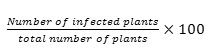
Rose plants were randomly inspected for the assessment of crown gall disease using disease incidence and severity levels. Disease incidence was determined using the formula by.10
Disease incidence =
While severity was assessed based on the size of the infection where mild; 1-3cm, moderate;4-6cm and severe; above 6cm.
Characterization of Agrobacterium tumefaciens
Biochemical Test
Biochemical test for the distinctive isolates was carried out following the Bergey’s manual of Determinative Bacteriology.10 Motility, gram stain, catalase, oxidase production, utilization of lactose, mannitol and salt tolerance (2%) tests were conducted.
Gram Staining
Gram staining was done by making bacterial smears from two days old cultures on sterile microscope slides. The smears were air-dried and then after, heat fixed by passing the slides over a flame and then Gram stained. The slides were observed under a compound microscope at magnification of × 1000.
Utilization of Lactose
Phenol red mannitol broth with 1% lactose was used. The broth contains peptone, phenol red(indicator) a Durham tube and carbohydrate(lactose). Each test tube was aseptically inoculated using an inoculating loop and incubated at 37 oC for 24 hours. A positive result was indicated by gas production (bubbles) on inverted Durham tube.
Motility Test
A semi solid agar medium was prepared in a test tube and inoculation done by use of a wire loop to make a single stab at the centre of the tube. Incubation at 37o C (conditions favoring motility) were carried out and examined at intervals (6, 24 and 48hours).
Growth on MacConkey Agar
Isolates were introduced into agar plates containing MacConkey agar by use of a sterile wire loop. The agar is selective and differential medium that is designed to isolate and differentiate gram-negative bacteria based on their ability to ferment lactose. Pink colonies on the MacConkey agar confirmed the bacteria to be gram negative.
Salt Tolerance
The nutrient broth was prepared by use of sodium chloride (65g) (1, 2, 3, 4, and 5%) and dextrose (1.0g) and allowed to settle at room temperature. Two to three colonies of the isolates were picked and inoculated into the broth and incubated. Turbidity was observed after 12 and 24 hours.
Oxidase Production
A filter paper was soaked with the substrate (tetramethyl-p-phenylenediamine dihydrochloride) and moistened with sterile distilled water. The bacterial colony was then picked using a wooden loop and smeared on the filter paper. Appearance of deep-purple colour was observed after 15 seconds.
Catalase Test
Slide method test was used for catalase test. Small amount of bacterial colony was transferred to the surface of a clean, dry glass slide by use of a sterile wire loop and a drop of 3% hydrogen peroxide added onto the slide and mixed. The rapid elaboration of oxygen bubbles after five seconds confirmed the bacterium catalyzes the breakdown of oxygen from hydrogen peroxide.
Pathogenicity Test
Isolates suspected to be A. tumefaciens were inoculated onto an indicator carrot disc bioassay and potato disc bioassay10. The same was inoculated onto healthy rose plants and observed.
Carrot Disc Assay
Carrots were obtained from a local market in Rongai. The collected carrots were then sterilized with 1-2% sodium hypochlorite and rinsed in three changes of sterile distilled water. Aseptically, the discs were prepared by chopping into smaller discs using a sterile surgical blade. The discs were placed in sterile petri dishes lined with sterile filter paper. Soriful procedures were followed and one hundred microliters of the inoculum was overlaid on each disc and sealed with parafilm and incubated in growth chamber at 280C for two weeks with continuous observation for tumor formation from meristematic tissues around the vascular system.
Potato Disc Assay
Potatoes were obtained from local market in Rongai. The collected potatoes were sterilized with 1-2% sodium hypochlorite and rinsed in three changes of sterile distilled water. Aseptically, the discs were prepared by chopping into smaller discs using a sterile surgical blade. The discs were placed in petri dishes containing water agar. According to10 recommendations, each disc was overlaid with 100µL of the inoculum and sealed with parafilm and incubated in growth chamber at 280C for two weeks with continuous observation. The discs were then stained with 5% Lugols Iodine (Iodine 5%, and potassium iodide 10%) for thirty (30) minutes and examined under microscope for tumor formation, where there was tumor formation, the starch was utilized.
Quantitative data was analyzed using statistical analysis software Genstat Discovery Edition 15 (VSN International Ltd. 2015) while qualitative data was assessed visually ant through the microscope.
Treatment means for quantitative data were compared using revised LSD test at the 0.05 level of probability using Fisher’s protected LSD.
Results
Effect of Amino Oligosaccharins and Alternaria Fine Protein on Incidence and Severity of Crown Gall
Disease severity was categorized into mild, moderate and severe (Table 1). Rose plants treated with various rates of amino oligosaccharins and alternaria fine protein had mild severity rates compared with the control plots in both sites. The incidence of the disease was assessed fortnightly for ten weeks (Table 2). In Winchester, there was no significance differences in plots applied with amino oligosaccharin and alternaria fine protein at the rate of 1.5g/L and plots applied with copper based fungicide. These plots showed a significant reduction in number of galls after the tenth week. In Bahati however, plots treated with amino oligosaccharin at 1.5g/L showed reduced number of galls on the tenth week after application compared with other treatments. The least incidence was observed in control plots in both farms.
The incidence and severity of the disease was assessed in each plot and control plots had the greatest incidence. Based on the progress of the disease after ten weeks of assessment, the least incidence was reported on plots treated with amino oligosaccharin and alternaria fine protein at 1.5g/L and were not significant different from the plots treated with copper based fungicides. The severity index shows infection being severe in control plots after ten weeks of assessment.
Table 1: Severity of crown gall affected by various treatments application
|
Treatment |
Disease Severity |
|||
|
Mild |
Moderate |
Severe |
||
|
Winchester |
||||
|
Amino oligosaccharins 0.5g/L |
69.0bc |
14.8b |
14.8cd |
|
|
Amino oligosaccharins 1g/L |
59.1c |
19.7a |
19.7cd |
|
|
Amino oligosaccharins 1.5g/L |
83.7a |
9.9c |
4.9d |
|
|
Copper at 2mL/L |
93.6a |
4.9d |
4.9d |
|
|
Control |
9.9e |
14.8b |
73.9a |
|
|
Bahati |
||||
|
Amino oligosaccharins 0.5g/L |
57.1c |
19.7a |
19.7cd |
|
|
Amino oligosaccharins 1g/L |
64.0bc |
9.9c |
24.6c |
|
|
Amino oligosaccharins 1.5g/L |
78.8ab |
4.9d |
14.8cd |
|
|
Copper at 2mL/L |
83.7a |
14.8b |
4.9d |
|
|
Control |
29.6d |
14.8b |
54.2b |
|
|
Mean |
63.1 |
12.8 |
23.6 |
|
|
LSD (P≤0.05) |
18.6 |
3.8 |
16.3 |
|
|
CV (%) |
25.9 |
5.3 |
22.8 |
|
Means followed by the same letters along the columns are not significant different at 5% level of probability
Table 2: Disease incidence progress as affected by various treatments application
|
Treatments |
Average number of galls |
||||
|
Pre- Treatment |
2 weeks |
4 weeks |
6 weeks |
10 weeks |
|
|
Winchester |
|||||
|
Amino oligosaccharins 0.5g/L |
1.4f |
1.5e |
1.4g |
2.1bc |
3.3a |
|
Amino oligosaccharins 1g/L |
2.1de |
1.9d |
1.5fg |
1.3d |
1.2bc |
|
Amino oligosaccharins 1.5g/L |
3.4a |
1.9d |
1.7ef |
1.3d |
1.1c |
|
Copper at 2mL/L |
2.5cd |
2.0d |
2.0cd |
2.0c |
1.9b |
|
Control |
1.5f |
1.5e |
1.9de |
2.4ab |
3.6a |
|
Bahati Amino oligosaccharins 0.5g/L |
2.0e |
2.6ab |
2.3ab |
2.1bc |
1.9b |
|
Amino oligosaccharins 1g/L |
2.3de |
2.1cd |
2.2bc |
1.5d |
1.0c |
|
Amino oligosaccharins 1.5/L |
2.9bc |
2.2cd |
2.0cd |
1.5d |
1.0c |
|
Copper at 2mL/L |
3.3ab |
2.8a |
2.0cd |
1.6d |
1.4bc |
|
Control |
2.0e |
2.4bc |
2.5a |
2.5a |
3.3a |
|
Mean |
2.34 |
2.09 |
1.95 |
1.83 |
1.97 |
|
LSD (P≤0.05) |
0.49 |
0.31 |
0.24 |
0.31 |
0.74 |
|
CV (%) |
0.4 |
0.16 |
0.1 |
0.17 |
0.97 |
Means followed by the same letters along the columns are not significant different at 5% level of probability
Effect of Amino Oligosaccharins and Alternaria Fine Protein on the Galling and Size of Galls
There were significant differences in number of galls in each treatment and in the two sites (P≤0.05) (Table 3). Application of the different rates of amino oligosaccharins had greater effect on the number of galls in Bahati than in Winchester. There was an increase in the number of galls in all the treatments in the second and fourth week after treatment application. However, decline in the number of galls was noticed sixth week after treatment application and it was least in plots treated with amino oligosaccharins at the rate of 1.5g/L. Application of amino oligosaccharins at 1.5g/L had greater effect on galling formation reducing the number significantly from 2.49 in the fourth week to 1.40 in the eight week and to 1.08 in the tenth week in Winchester while in Bahati, the number reduced significantly from 1.54 to 1.03 ten weeks after treatment application. The results were not significant different from the plots treated with copper at 2mL/L in both farms. However, control plots had the number of galls increasing every week in both farms.
Table 3: Effect of amino oligosaccharins at different rates on the number of galls induced by A. tumefaciens in Winchester and Bahati
|
Treatment |
Weeks after treatment application |
||||
|
2 |
4 |
6 |
8 |
10 |
|
|
Winchester |
|||||
|
Amino oligosaccharins 0.5g/L |
1.99abcd |
1.92de |
2.05d |
2.48b |
2.56ab |
|
Amino oligosaccharins 1g/L |
2.38ab |
2.78ab |
2.58bc |
2.48b |
2.29bc |
|
Amino oligosaccharins 1.5g/L |
2.11abc |
2.49bc |
2.18cd |
1.40ef |
1.08d |
|
Copper at 2mL/L |
2.12abc |
2.42bc |
2.21cd |
1.50def |
1.38cd |
|
Control |
2.46a |
3.12a |
3.12a |
3.24a |
3.30a |
|
Bahati |
|||||
|
Amino oligosaccharins 0.5g/L |
1.54de |
1.58e |
1.54e |
2.17bcd |
2.67ab |
|
Amino oligosaccharins 1g/L |
1.87bcde |
2.04cd |
2.00d |
2.00bcde |
2.43ab |
|
Amino oligosaccharins 1.5g/L |
1.37e |
1.54e |
1.46e |
1.25f |
1.03dcd |
|
Copper at 2mL/L |
1.67cde |
1.50e |
1.46e |
1.75cdef |
2.56ab |
|
Control |
1.92bcd |
1.83de |
2.79ab |
2.42bc |
3.17ab |
|
Mean |
1.67 |
1.70 |
1.85 |
1.92 |
2.43 |
|
LSD (P≤0.05) (Trt × Site) |
0.51 |
0.47 |
0.44 |
0.71 |
1.00 |
|
CV (%) |
18.00 |
15.1 |
15.60 |
22.30 |
25.00 |
Means followed by the same letters along the columns are not significant different at 5% level of probability
There were significant differences in the size of galls before and after treatment applications (Table 4). There was significant reduction in sizes of galls after treatment application with the highest percentage reduction observed in plots treated with amino oligosaccharins at the rate of 1.5g/L and amino oligosaccharins at the rate of 1g/L in both sites. Application of copper fungicide as the standard also resulted in reduced gall sizes in both sites. There was significant increase in gall sizes in control plots.
Table 4: Effect of amino oligosaccharins at different rates on the size of galls induced by A. tumefaciens in Winchester and Bahati
|
Treatment |
Gall size before treatment application (mm) |
Gall size 10 wks. after treatment application (mm) |
% Reduction in size |
|
Winchester |
|||
|
Amino oligosaccharins 0.5g/L |
10.0 |
9.3bc |
7.0c |
|
Amino oligosaccharins 1g/L |
12.2 |
8.6bcd |
29.5abc |
|
Amino oligosaccharins 1.5g/L |
14.0 |
7.6cd |
45.7a |
|
Copper at 2mL/L |
7.9 |
6.2d |
21.5bc |
|
Control |
12.0 |
16.4a |
-36.7d |
|
Bahati |
|||
|
Amino oligosaccharins 0.5g/L |
11.3 |
10.2b |
9.7bc |
|
Amino oligosaccharins 1g/L |
13.2 |
10.3b |
22.0bc |
|
Amino oligosaccharins 1.5g/L |
12.6 |
8.7bcd |
31.0ab |
|
Copper at 2mL/L |
8.9 |
7.4cd |
16.9b |
|
Control |
9.3 |
14.6a |
-57.0d |
|
Mean |
11.1 |
9.7 |
8.9 |
|
LSD (P≤0.05) |
1.4 |
2.5 |
22.7 |
|
CV (%) |
4.1 |
12.4 |
33.8 |
Means followed by the same letters along the columns are not significant different at 5% level of probability
Biochemical Tests
The biochemical tests for the different isolates of A. tumefaciens are presented in Table 5. The gram reaction indicates that the selected isolates were gram negative. The isolates were gram positive for motility, catalase, oxidase, lactose, mannitol, and salt tolerance tests. In terms of shape, the colonies were circular and slightly raised and were cream white in colour with smooth margins (Table 6)
Table 5: Characteristics of the selected strains of A. tumefaciens
|
Biochemical tests |
Bahati |
Winchester |
||||||
|
Bht 1 |
Bht 2 |
Bht 3 |
Bht 4 |
Krn 1 |
Krn 2 |
Krn 3 |
Krn 4 |
|
|
Gram stain |
– |
– |
– |
– |
– |
– |
– |
– |
|
Motility Test |
+ |
+ |
+ |
+ |
+ |
+ |
+ |
+ |
|
Catalase test |
+ |
+ |
+ |
+ |
+ |
+ |
+ |
+ |
|
Oxidase test |
+ |
+ |
+ |
+ |
+ |
+ |
+ |
+ |
|
Lactose |
+ |
+ |
+ |
+ |
+ |
+ |
+ |
+ |
|
Mannitol |
+ |
+ |
+ |
+ |
+ |
+ |
+ |
+ |
|
Salt tolerance |
+ |
+ |
+ |
+ |
+ |
+ |
+ |
+ |
+: Positive, –: Negative, Bht 1: Bahati sample 1, Krn 1: Karen sample 1
Table 6: Morphological characteristics of A. tumefaciens
|
Character |
Nutrient Agar |
|
Shape |
Circular, slightly raised |
|
Color |
Cream white, |
|
Surface margin |
Smooth |
Pathogenicity Test for Agrobacterium tumefaciens Isolates
A.tumefaciens isolates from both farms produced tumors when inoculated on carrot and potato discs. Young galls (tumors) were observed developing at the central part of the carrot and potato discs two weeks after inoculation. However, no symptoms were observed in control discs.
Relationship among Flower Growth Parameters and Crown Galls
From correlation analysis, significant positive correlation was displayed between total number of galls and the size of gall (0.6416, P≤ 0.05). However, there was a significant negative correlation between the number of galls and shoot length (-0.695, P≤ 0.05).
Table 7: Correlation coefficient among flower growth parameters and crown galls
|
|
Shoot length |
Size of galls |
Stem length |
Total galls |
|
Shoot length |
– |
|||
|
Size of galls |
0.0193 |
– |
||
|
Stem length |
0.4628 |
-0.1858 |
– |
|
|
Total galls |
-0.695* |
0.6416* |
-0.0809 |
– |
Discussion
Application of amino oligosaccharins had greater effect on the number of galls; however, the effect depended on the rate of application. There was increase in the number of galls in all the treatments in the second and fourth week after treatment application. However, reduction in the number of galls was noted in the sixth week after treatment application and it was highest in plots treated with amino oligosaccharins at the rate of 1.5g/L. Amino oligosaccharins and alternaria fine proteins can constitute an important control agent for crown gall in roses. Similar results were reported by.11 In their findings,11 found chitosan and its derivatives to inhibit the growth of A. tumefaciens and Erwinia spp and displayed highest antibacterial activity against A. tumefaciens, Erwinia spp with MIC 500 mg/L and 480 mg/L, respectively. Similarly, when oligosaccharin derivatives chitosan were used in controlling Xanthomonas vesicatoria, infections were greatly reduced by above 60%12, reported significant reduction of the disease incidence and delayed symptoms development and the size of the lesions were smaller in comparison with other treatments.
The reduction of number of galls was due to oligosaccharides derivatives unique elicitation of physiological and biochemical changes in roses resulting to induced resistance.11 There is an enhanced activity of enzymes that are linked to defense against plant diseases in response to application of oligosaccharins. Enzymes such as Chi, β-1,3-Glucanase(GLU), peroxidase (PO), phenylalanine ammonia-lyase (PAL) and polyphenol oxidase (PPO) are activated and are able to eliminate infectious agents such as Agrobacterium tumefaciens.12 Increase in Phenylalanine ammonia-lyase (PAL) enzyme activity is important as it manages pathogenic infection.13 working on tomatoes reported enhanced activities in response to Ralstonia solanacearum inoculation in tomato pre-treated with chitosan.
According to 14,12 oligosaccharins act by inducing several activities of defense enzymes in roses that are involved in pathogen defense. Alternaria fine protein accelerates plant growth vitality, increase proline content, strengthen cellulase and promote the growth of plant cells, strengthen root activity, thereby managing plant diseases. Chitosan, a biopolymer amino polysaccharides has been reported to have strong antibacterial activities against plant pathogenic bacteria such as A. tumefaciens, Erwinia spp,11 Streptomyces scabies,15 Xanthomonas spp,16 Pseudomonas syringae,17 However, the activity of chitosan oligosaccharins depends on the concentration11,18and molecular weight 19and the type of bacteria.20
The mechanism of antimicrobial activity of chitosan and its derivatives remains a mystery as it has not been understood well and it is therefore considered to be complex. However, according to,21 at the initial stages chitosan ties with negatively charged components on the bacterial surface by means of electrostatic interactions which changes the permeability of bacterial wall and permits chitosan to get to the internal cell targets subsequently closing down cell division causing death.22
In this study, bacteria were isolated using nutrient agar media which has been previously used for isolation of A. tumefaciens from rose samples with crown gall. A series of biochemical tests such as motility, gram staining, oxidase production and catalase, utilization of lactose and mannitol, salt tolerance (2%) potato and carrot disc assay revealed that the isolated bacterium was gram negative and had the capacity to cause tumor in plant disc sample.
The biochemical approaches have been utilized in the past investigations for the recognizable proof of A. tumefaciens from crown gall samples from diverse plant species.14 Tumor forming ability of the isolates confirmed that they were virulent, however, according to 25, virulence of A. tumefaciens was because of the nature of the host, internal physiology of the strains and environmental conditions. Crown gall disease can be severe in rose plantations if young trees are infected. A. tumefaciens was found on root surfaces and was effectively isolated from galls. The bacterium was isolated and confirmed using different morphological and biochemical tests.
Conclusion
The study demonstrates the ability of amino oligosaccharins and alternaria fine proteins to be used as an alternative to management of plant diseases. A significant reduction in the number of galls and size following application of amino oligosaccharins and alternaria fine proteins was observed as well as improvement in plant growth. The ability of the treatments to manage the disease can be attributed to enhanced defense enzyme activity. Therefore, this study provides good support for incorporation of amino oligosaccharins and alternaria fine proteins in the management and yield enhancement of roses.
Acknowledgements
Special acknowledgements go to Higher Education Loans Board(HELB) post graduate scholarship program that awarded me a scholarship that catered for part of my fees, may you never lack so that you continue assisting others in need.
I wish to acknowledge also the contribution of CCIPT Director Mr. Fan Liqiang who provided the samples that were used in this research.
References
- Rikken, W. 2011.The global competiveness of the Kenyan flower industry world bank. Retrieved March 2, 2013. Available: http://www.Kenyaflowercouncil.org/pdf/vc5520global%20competiveness%20kenyan%20flower%20industry%20%-%20proverde.pdf.
- Patton, D. (2008). Kenya: Fair-trade Gives Flowers an Edge in UK. Business Daily (Nairobi).
- Murigi, J.G. 2010.Management of crown gall disease of roses using Agrobacterium radiobacter, corn oil and copper hydroxide and oxychloride in Kenya. MSc. Thesis, Kenyatta University.
- Muhammad, A. (2009). Would African Countries Benefit from the Termination of Kenya’s Economic Partnership Agreement (EPA) with the EU? An Analysis of EU Demand for Imported Roses. Journal of Agricultural Economic, 60 (1): 220-238.
- HCDA, 2010. Horticultural Crops Development Authority: Volumes and Values Export Statistics. Nairobi, Kenya.
- Lorenzo, H., Cid, M. C., Siverio, J. M., Ruano, M. C. (2000). Effects of sodium on mineral nutrition in rose plants. Annuals of Applied Biology, 139: 567
- Deacon, J. 2002.Biology and control of crown gall. The microbial world pp12. http:/helios.bto.ed.ac.uk/bto/microbes/crown.htm.
- Rouhrazi, K., Rahimian, H. 2014. Biochemical and genetic characterization of Agrobacterium tumefaciens the causal agent of walnut crown gall disease in Iran. Archives of Phytopathology and Plant Protection. 47:20, 2493-2500, DOI: 10.1080/03235408.2014.880575
- Tolba, I.H., Soliman, M.A. 2013. Efficacy of native antagonistic bacterial isolates in biological control of crown gall disease in Egypt. Annals of Agricultural Science. 58(1), 43–49
- Holt, J. G., Kreig, P.H.A., Sneath, J.T. S., Williams, S.T. 1994. Bergey’s manual of determinative bacteriology. 9theditionWilliams and Wilkins Baltimore.
- Rabea E.I., Badawy M.E.I., Stevens C.V., Smagghe G., Steurbaut W. (2003): Chitosan as antimicrobial agent: applications and mode of action. Biomacro- molecules, 4: 1457–1465.
- Rabea, E.I. and Steurbaut, W, 2010. Chemically modified chitosans as antimicrobial agents against some plant pathogenic bacteria and fungi. Plant Protect. Sci., 46: 149-158
- Ramkissoon, A., Francis, J., Bowrin, V., Ramjegathesh, R., Ramsubhag, A., Jayaraman, J., 2016. Bio-efficacy of a chitosan based elicitor on Alternaria solani and Xanthomonas vesicatoria infections in tomato under tropical conditions. Annals of Applied Biology.169 274–283
- Mandal S., Kar I., Mukherjee A.K., Acharya P. 2013. Elicitor-induced defense responses in Solanum lycopersicum against Ralstonia solanacearum. The Scientific World Journal, 2013, 561056.
- El Hadrami, A., Adam, L.R., El Hadrami, I. and Daayf, F. 2010. Chitosan in plant protection. Marine Drugs 8:968-987.
- Beausejour, J., N. Clermont and C. Beaulieu, 2003.Effect of Streptomyces melanosporofaciens strain EF-76 and of chitosan on common scab of potato. Plant Soil, 256: 463-468.
- Li, B., X. Wang, R. Chen, W. Huangfu and G.L. Xie, 2008. Antibacterial activity of chitosan solution against Xanthomonas pathogenic bacteria isolated from Euphorbia pulcherrima. Carbohydrate Polymers 72: 287-292.
- Ferrante, P. and M. Scortichini, 2010. Molecular and phenotypic features of Pseudomonas syringae actinidiae isolated during recent epidemics of bacterial canker on yellow kiwifruit (Actinidia chinensis) in central Italy. Plant Pathology 59: 954-962.
- Yang, C., B. Li, M. Ge, K. Zhou and Y. Wang et al., 2014. Inhibitory effect and mode of action of chitosan solution against rice bacterial brown stripe pathogen Acidovoraxavenae avenae RS-1. Carbohydr. Res., 391: 48-54.
- Liu, N., X.G. Chen, H.J. Park, C.G. Liu, C.S. Liu, X.H. Meng and L.J. Yu, 2006. Effect of MW and concentration of chitosan on antibacterial activity of Escherichia coli. Carbohydrates. Polymers 64: 60-65.
- Shanmugam, A., K. Kathiresan and L. Nayak, 2016.Preparation, characterization and antibacterial activity of chitosan and phosphorylated chitosan from cuttlebone of Sepia kobiensis (Hoyle, 1885). Biotechnol. Rep., 9: 25-30.
- Raafat D., Bargen K., Haas A. (2008): Insights into the mode of action of chitosan as an antibacterial compound. Journal of Applied Environmental Microbiology, 74: 3764–3773.
- Je J.Y., Kim S.K. 2006: Chitosan derivatives killed bacteria by disrupting the outer and inner membrane. Journal of Agriculture and Food Chemistry, 54: 6629–6633
- China patent. 2013. Plant growth regulation composition containing plant activator protein. https://patents.google.com/patent/CN103828828A/en
- Karthy E. S., Ranjitha P., Mohankumar A. 2009. Antimicrobial potential of plant seed extracts against multidrug resistant methicillin resistant Staphylococcus aureus (MDR-MRSA). IJB 1:34-40
- Sarker, A.Q., Mondol, P.C., Islam, S., Alam, M. F. 2011. Identification of virulent Agrobacterium tumefaciens strains from some dicotyledonous plants in Bangladesh. Agriculturae Conspectus Scientificus. 76: (2)147-152)